
(vv)Anatomy Of Eye Movement.mp4(tt)
From: Root T. Ophthalmology Lecture - Tropias & Phorias (part 1/2) Retrieved from: https://www.youtube.com/watch?v=dRYBOBSyzAU

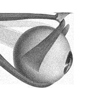
https://embryology.med.unsw.edu.au/embryology/index.php/File:Extraocular-muscles-scan.jpg
Elevation of the abducted eye is primarily accomplished by the superior rectus, while elevation of the adducted eye is primarily accomplished by the inferior oblique1.
Depression in abduction isolates the inferior rectus while depression in adduction isolates the superior oblique.
Note that the oblique muscles are primarily elevators and depressors in adduction.
A corollary of the above is evidenced by some of the various actions of the extra-ocular muscles:
- RAD: Rectus muscles ADduct: superior and inferior rectus muscles ADduct
- SIN: Superior muscles INtort: the superior rectus and superior oblique muscles INtort
It follows that the Inferior muscles EXtort the eye:
- The inferior rectus depresses the abducted eye and secondarily extorts the eye.
- The inferior oblique extorts the eye and secondarily elevates the adducted eye.
The obliques' tertiary action is ABduction: hence, with a superior oblique palsy, there is deviation inwards with downgaze .


From: Peterson E. The Motility Exam. Moran CORE. Available at http://morancore.utah.edu/basic-ophthalmology-review/the-motility-exam/
The range of eye movements is examined by asking the patient to follow a target, placed 50 cm away in order to avoid convergence2. Note that each cardinal position is primarily obtained by the action of one muscle1. Subjective diplopia in the absence of visible ocular motility restriction should be further tested by occluding each eye in turn, and the general rule is that the outer image comes from the paretic eye. For example, if the patient reports diplopia in left gaze and the outer image disappears with right eye occluded, the patient has a right medial rectus palsy2.
It is conventional to record cardinal eye positions in an H fashion from the patient’s perspective2.

From: Peterson E. The Motility Exam. Moran CORE. Available at http://morancore.utah.edu/basic-ophthalmology-review/the-motility-exam/
It is conventional that if there is a vertical deviation of the eyes, the higher of the two is referred to as hypertropic/hyperphoric, regardless of which eye is at fault.
THERE ARE THREE COVER TESTS2:
Detection of a subtle palsy is best performed using the alternate cover test in the six cardinal positions of gaze: Diagnosis of a subtle oculomotor palsy
(Note that the terms used for the various cover tests are those adopted by Leigh and Zee (the terms vary tremendously from one text to another))3.