
A sagittal shift forwards is a combination of1:
a) Retrocaput (extension at atlantooccipital joint)
b) Anterocollis
This may develop into a “Goose-neck” posture: an anterior sagittal shift on the cervical spine.
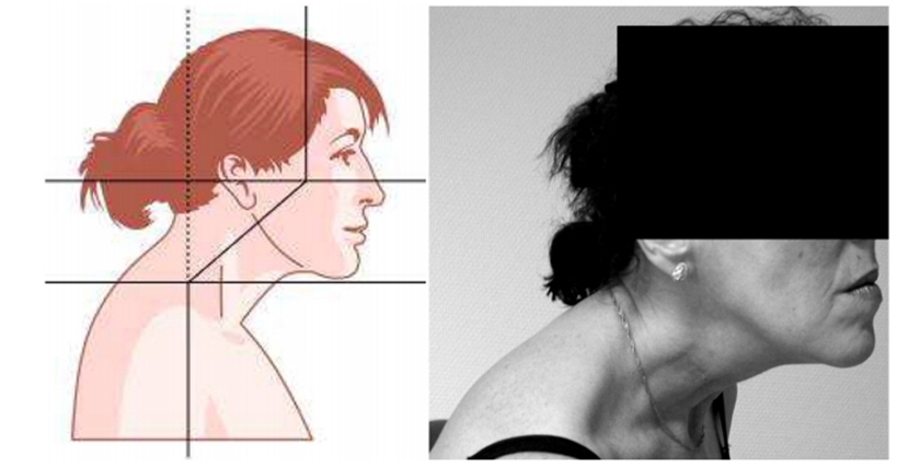
Normally, when looking straight ahead (see Figure 2), the head is aligned almost horizontally on the C-spine. There is a lordosis of the C-spine and hence a flexion of the C-spine on the T-spine. The longus colli and the posterior cervical muscles form a sleeve, which encloses and stabilizes the cervical spine in all positions of the head. The stabilization of the head and neck is also assisted by both the left and right levator scapulae muscles, which act like a pair of reins to hold the head upright and horizontally on the cervical spine. Thus, normally, the head pivots on the cervical spine, which we can be imagined as a ‘‘see saw’’ in a balanced position.
Simultaneous activation of both splenius capitii muscles and SCM muscles produces an extension of the head on the cervical spine, resulting in an extreme hyperlordosis of the C-spine. It appears that it is the synchronous activity of both groups of muscles (SCMs and splenius capitis) that gives rise to the goose-neck posture, as bilateral splenius activation alone causes extension of the head on the C-spine, resulting in retrocollis).
From: Reichel G. Dystonias of the Neck: Clinico-Radiologic Correlations [Internet]. In: Dystonia - The Many Facets. InTech; 2012. Available from: http://www.intechopen.com/books/dystonia-the-many-facets/dystonias-of-the-neck-clinico-radiologic-correlations
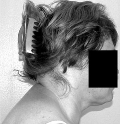
Shift of the head forwards. Even when supine, the hypertrophic sternocleidomastoid muscles are bilaterally tensed, and pull the head backwards and the cervical spine forwards.
From: Reichel G. Cervical dystonia: A new phenomenological classification for botulinum toxin therapy. Basal Ganglia 2011; 1: 5–12.
a) Retrocaput (extension at atlantooccipital joint)
b) Anterocollis

From: Reichel G. Dystonias of the Neck: Clinico-Radiologic Correlations [Internet]. In: Dystonia - The Many Facets. InTech; 2012. Available from: http://www.intechopen.com/books/dystonia-the-many-facets/dystonias-of-the-neck-clinico-radiologic-correlations
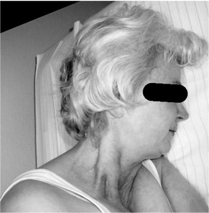
Figure shows goose neck posture with 1 indicating splenius capitis bilaterally, and 2 & 3 the sub-occipital muscles.
Sternocleidomastoid may also be active bilaterally2.
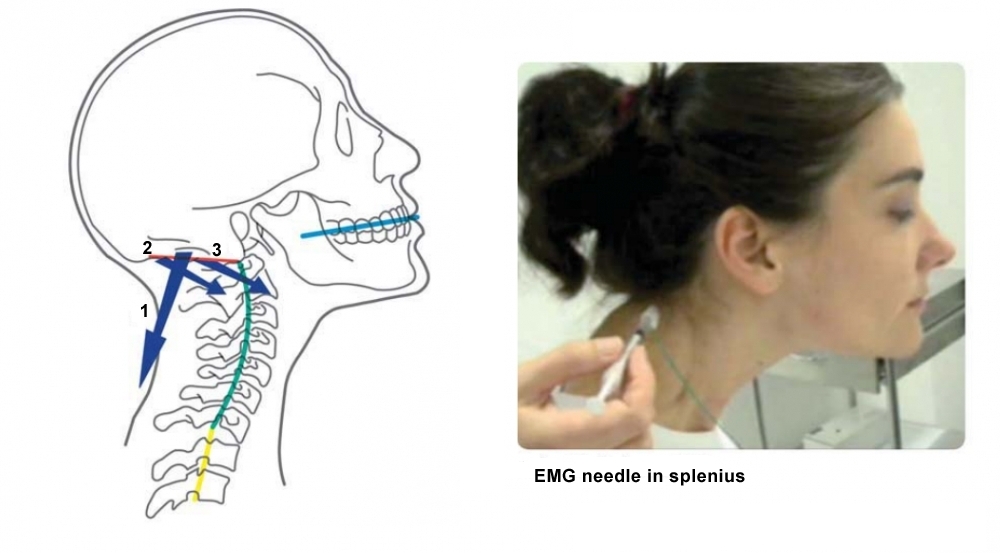
From: Flowers JM, Hicklin LA, Marion MH. Anterior and posterior sagittal shift in cervical dystonia: A clinical and electromyographic study, including a new EMG approach of the longus colli muscle. Mov Disord 2011; 26: 2409–14.
Forward sagittal shift typically involves the sternocleidomastoid and the splenius capitis muscles.
| WARNING: risk of dysphagia with injections |