LONGISSIMUS CAPITIS LONGISSIMUS CERVICIS RECTUS CAPITIS POSTERIOR MAJOR SEMISPINALIS CERVICIS SEMISPINALIS CAPITIS
-
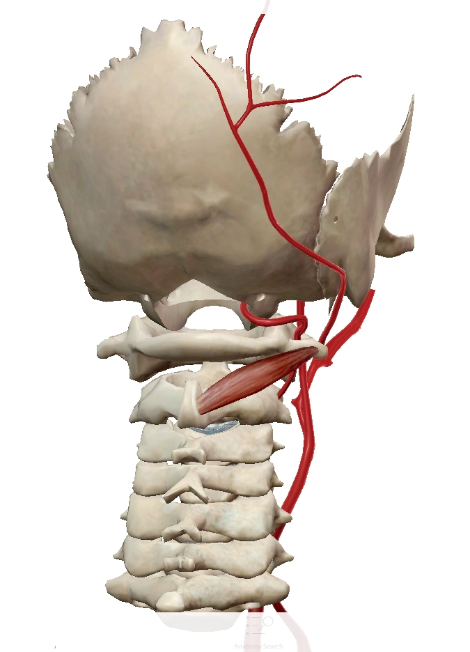
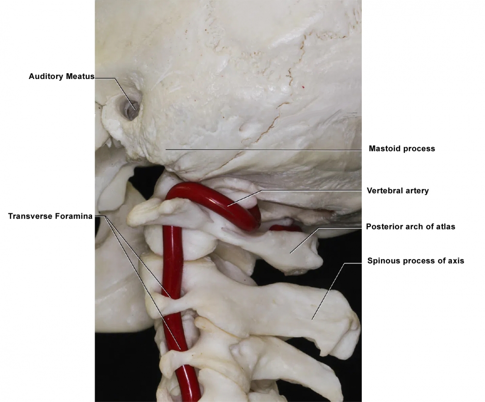
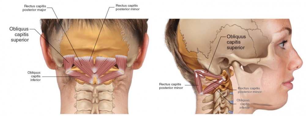
Origin: posterior spinous process and adjacent lamina of axis.
-
Insertion: transverse process of atlas
-
Action: rotates atlas upon axis, thereby rotating head to same side.
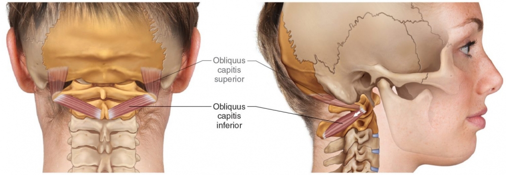
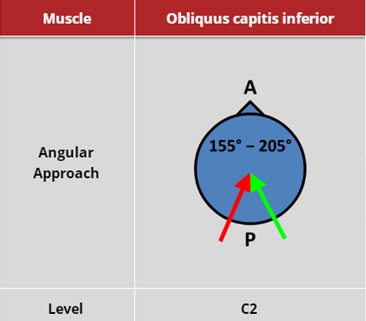
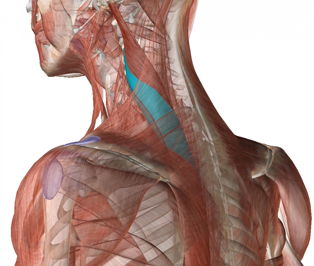
The almost horizontal obliquus capitis inferior (OCI) is inserted on the transverse process of the atlas and originates from the spinous process of the axis (C2). It contributes to head movement related to fast visual exploration.
OCI is the biggest suboccipital muscle and is the 2nd strongest head rotator (after the sternocleidomastoid).
The depth from the skin to the superficial fascia of the muscle ranges from about 10 mm to over 35 mm. The muscle thickness also has a wide degree of variability, ranging from 10 to 20 mm, and when it contracts, its thickness increases and the length shortens.
From: Dr. Joe Muscolino. www.learnmuscles.com – art work Giovanni Rimasti
 |
 |
 |
 |
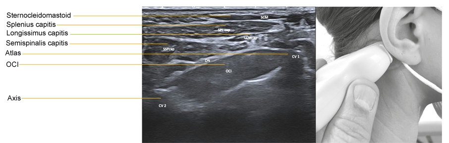
From: Fietzek UM, Nene D, Schramm A, Appel-Cresswell S, Košutzká Z, Walter U, Wissel J, Berweck S, Chouinard S, Bäumer T. The Role of Ultrasound for the Personalized Botulinum Toxin Treatment of Cervical Dystonia. Toxins (Basel). 2021 May 20;13(5):365.
Three different approaches to injection of OCI
A: In plane technique
B: Out of plane technique
C: Sagittal approach: out of plane approach.
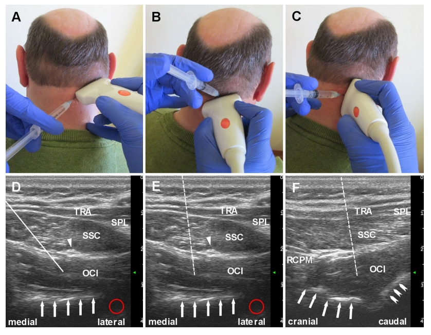
TRA: Trapezius; SPL: Splenius; SSC: Semispinalis capitis; RCPM: Rectus capitis posterior major
White arrows indicate bone; red circle indicates possible site of vertebral artery. Triangle: Greater occipital nerve
Dashed line (E and F) indicates potential path of the needle
From: Walter U, Dudesek A, Fietzek UM. A simplified ultrasonography-guided approach for neurotoxin injection into the obliquus capitis inferior muscle in spasmodic torticollis. J Neural Transm (Vienna). 2018 Jul;125(7):1037-1042.
(vv)OCI.mp4(tt)
(vv)OCIfietz.mp4(tt)
Segment 1: The initial positioning of the ultrasound probe at the patient’s neck is demonstrated. After visualization of the processus spinosus of vertebra C4 the ultrasound probe is shifted in cranial direction in order to display the OCI at the level of vertebra C1. Segment 2: For full display of the OCI muscle on axial imaging plane the ultrasound probe is shifted in medio-lateral direction and slightly twisted, moving the anterolateral part in an upwards direction.
Segment 3: For optimal display of the OCI muscle belly on sagittal imaging plane the ultrasound probe is rotated by 90 degrees.
Segment 4: The US-guided insertion of the injection needle (here connected to an electromyography machine) is presented. The needle tip is visible on the screen. Note the electromyography noise demonstrating dystonic activity of the OCI muscle. During the injection of the BoNT the electromyography noise disappears.
From: Walter U, Dudesek A, Fietzek UM. A simplified ultrasonography-guided approach for neurotoxin injection into the obliquus capitis inferior muscle in spasmodic torticollis. J Neural Transm (Vienna). 2018 Jul;125(7):1037-1042.
-
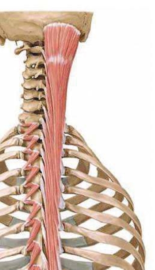
Origin: posterior surface of transverse processes of T1 to T6
-
Insertion: posterior margin of mastoid process and the temporal bone. The insertion is deep to that of the sternocleidomastoid and splenius muscles.
-
Action: Acting bilaterally, extends and hyperextends head
Acting unilaterally, flexes and rotates the head ipsilaterall (ipsiversion). The rotation is ipsilateral, and rotation of the head takes place at the atlanto-axial joint.
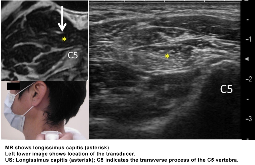
From: Mezaki T. Ultrasoundâ€guided botulinum toxin therapy for deep muscles in cervical dystonia. Neurol Clin Neurosci 2020; 8: 3–10.

From: Fietzek UM, Nene D, Schramm A, Appel-Cresswell S, Košutzká Z, Walter U, Wissel J, Berweck S, Chouinard S, Bäumer T. The Role of Ultrasound for the Personalized Botulinum Toxin Treatment of Cervical Dystonia. Toxins (Basel). 2021 May 20;13(5):365.
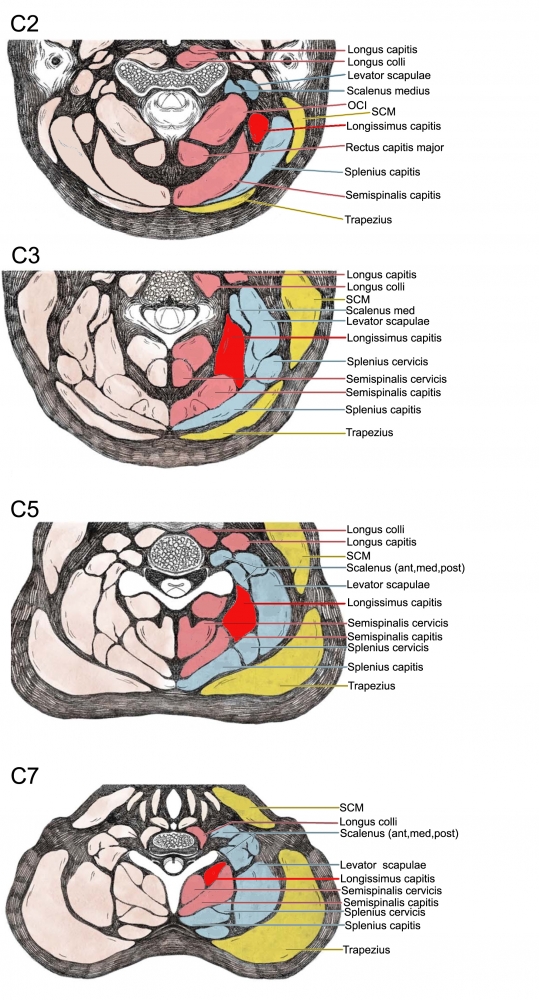
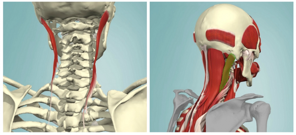
Note that in the figure on the right, splenius capitis is transparent, allowing for the longissimus capitis to be seen deep to splenius.
The longissimus capitis lies between the longissimus cervicis (which is directly lateral to it) and the splenius capitis (which is directly medial to it).
 |

-
Origin: Transverse processes of the first 5 thoracic vertabrae
-
The transverse process attachment of the levator scapulae is anterior to the transverse process attachment of the splenius cervicis and directly superficial (posterior) to the transverse process attachment of the scalenes.
-
Insertion: Posterior tubercles of the transverse processes of C2-C5
-
Action: Bilateral contraction leads to extension of the neck.
Unilateral contraction causes lateral flexion and rotation of the head ipsilaterally
From: Fietzek UM, Nene D, Schramm A, Appel-Cresswell S, Košutzká Z, Walter U, Wissel J, Berweck S, Chouinard S, Bäumer T. The Role of Ultrasound for the Personalized Botulinum Toxin Treatment of Cervical Dystonia. Toxins (Basel). 2021 May 20;13(5):365.

-
Origin: posterior tubercle of atlas.
-
Insertion: medial portion of occipital bone below inferior nuchal line.
-
Action: extends head.
Rotates head to same side.
-
Origin: Transverse processes of T1-T6
-
Insertion: Spinous processes of C2-C5
-
Action: Bilateral contraction leads to extension of the neck.
Unilateral contraction causes lateral flexion ipsilaterally and contralateral rotation
 |
 |

From: Fietzek UM, Nene D, Schramm A, Appel-Cresswell S, Košutzká Z, Walter U, Wissel J, Berweck S, Chouinard S, Bäumer T. The Role of Ultrasound for the Personalized Botulinum Toxin Treatment of Cervical Dystonia. Toxins (Basel). 2021 May 20;13(5):365.
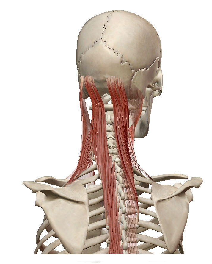
-
Origin: transverse processes of the lower four cervical and upper six or seven thoracic vertebrae (C4–T7).
-
Insertion: between the superior and inferior nuchal lines of the occipital bone.
-
Action: Bilateral contraction leads to extension of the neck.
Unilateral contraction causes contralateral rotation
 |
 |
 |
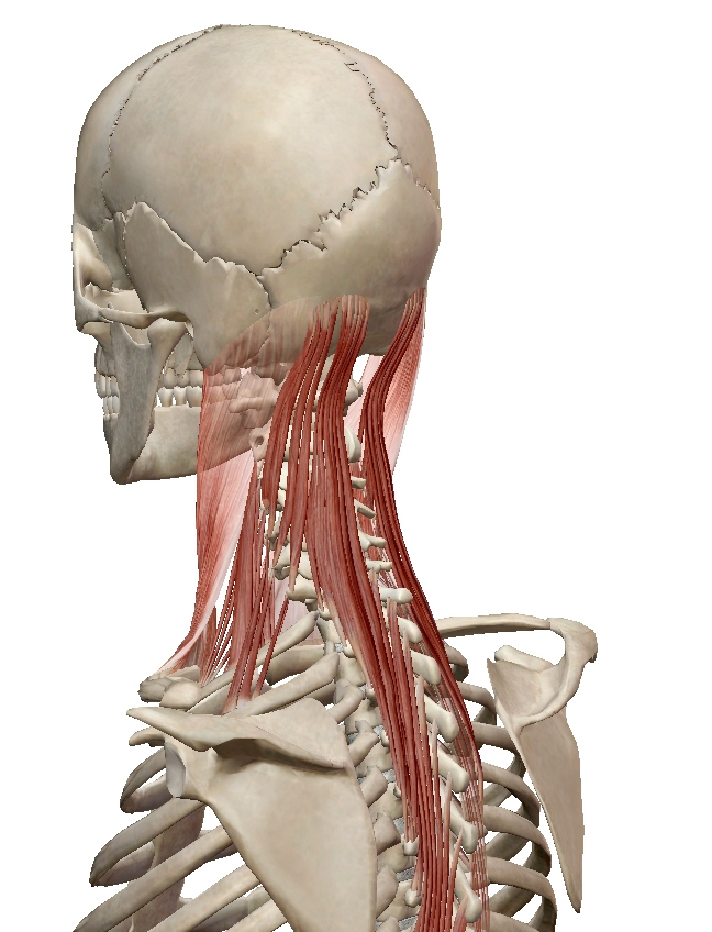 |
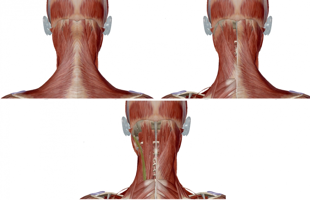
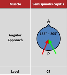
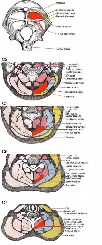
Figure 12. Trajectory of approach and relationships of the surrounding muscles to Semispinalis capitis
 |
 |
From: Fietzek UM, Nene D, Schramm A, Appel-Cresswell S, Košutzká Z, Walter U, Wissel J, Berweck S, Chouinard S, Bäumer T. The Role of Ultrasound for the Personalized Botulinum Toxin Treatment of Cervical Dystonia. Toxins (Basel). 2021 May 20;13(5):365.
(vv)semispinalis capitis.mp4(tt)
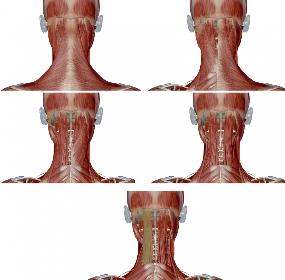
1. Trapezius and sternocleidomastoid muscles removed
2. Splenius removed.
3. Longissimus capitis and cervicis removed.
4. Levator scapulae removed showing semispinalis capitis.