
-
Origin:
Sternal Head: upper part of anterior surface of manubrium sternum
Clavicular Head: upper surface of medial third of clavicle -
Insertion:
Sternal Head: lateral one half of superior nuchal line of occipital bone
Clavicular Head: outer surface of mastoid process -
Action:
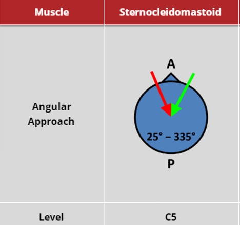
Unilateral contraction: rotates head to opposite side (contraversion); flexes head to same side
Bilateral contraction: draws head forwards (protracts) and brings about anterior flexion of the neck.
In retroflexion, may act to further exacerbate retroflexion.
 |
 |
 |
 |
(vv)SCM without US.mp4(tt)
(vv)SCM.mkv(tt)
-
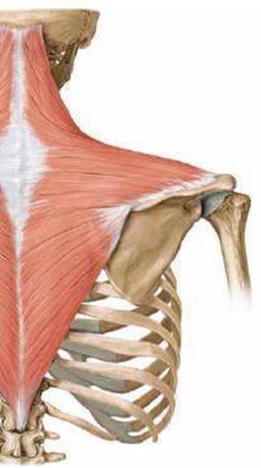
Origin: medial third of superior nuchal line of occipital bone.
External occipital protuberance and ligamentum nuchae.
Spinous processes and supraspinous ligaments of seventh cervical vertebra (C7) and all thoracic vertebrae (T1–12) -
Insertion: .Superior edge of crest of spine of scapula and the medial border of acromion.
Posterior border of lateral one-third of clavicle. -
Action: powerful elevator of the scapula; rotates the scapula during abduction of humerus above horizontal.
Middle fibers retract scapula.
Lower fibers depress scapula, particularly against resistance, as when using hands to get up from a chair.
Extension of neck
Contraversion of neck
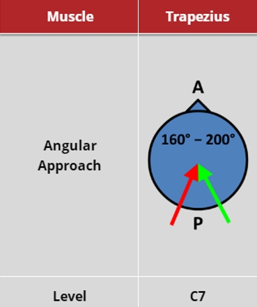
Trapezius 2 x 7.5-10 u Max 50 u Botox; 2 x 30-40 max 200 u Dysport
The trapezius is superficial and is generally easily palpated at the base of the neck. The muscle is usually considered to have three functional parts: upper, middle, and lower. The trapezius’ lateral attachments are the same as the proximal attachments of the deltoid: the lateral clavicle, the acromion process, and the spine of the scapula.
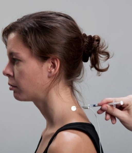
The trapezius has a variable role in causing abnormal head posture, but, at the least, the muscle often seems to cause pain, which is effectively treated with botulinum toxin.
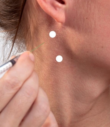
Recommended to inject trapezius at the base of the neck with one or two injections.
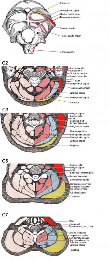
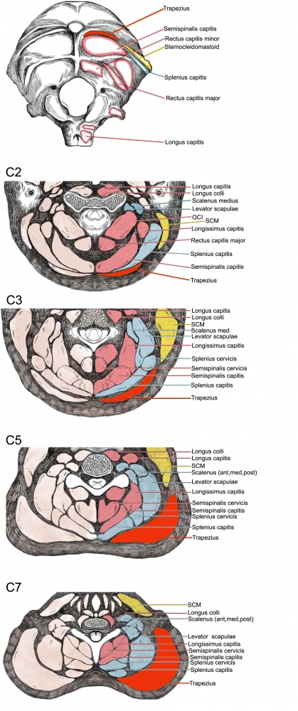
Relations: directly deep to the trapezius in the neck are the semispinalis capitis, the splenius capitis, and the levator scapulae.
Directly anterior to the anterior border of the trapezius are the splenius capitis, the levator scapulae, and the scalenes (in the posterior triangle of the neck)
 |
 |
 |
 |
The video shows positioning of the ultrasound probe and of the EMG needle. The muscle belly is visible on the background screen.
(vv)Trapezius.mp4(tt)