MYELOPATHY

INDEX


NMOSD            MOG Ab Disease               Sarcoidosis         Paraneoplastic           Dural AVF            Cord Infarction           Cervical Spondylosis

References


Mulitple Choice Self Test

Q1: Patient with an acute myelopathy. What is seen in the axial T2 weighted images?.
    a: Bright Spotty Lesions.
    b: Transverse myelitis.
    c: Central cord necrosis.
    d: Cord infarction.
Q2: The findings in the previous question are most suggestive of?.
    a: CMV infection.
    b: MOG.
    c: NMOSD.
    d: Neurosyphilis.
Q3: This 27 year old woman presented with acute paraplegia. What do the MRI findings suggest? (Sagittal T2 weighted image).
    a: Syringomyelia.
    b: Ependymoma.
    c: Transverse myelitis.
    d: Hyperintense swelling at cervical and thoracic levels .
Q4: What is the likely cause of the imaging findings seen in the previous question? (Sagittal T2 weighted image).
    a: MOG antibody disease.
    b: Neurosyphilis.
    c: NMOSD .
    d: Paraneoplastic .
Q5: A 31-year-old man presented with acute onset of paraparesis, numbness with T8 sensory level, and urinary retention. Sagittal T2-weighted MRI shows?.
    a: Artefact.
    b: Diffuse longitudinal signal change.
    c: Normal.
    d: Features of TB myelomeningoradiculapathy.
Q6: The most likely cause of the patient's problem is?.
    a: Bilharzia of spinal cord.
    b: Echinococcal cysts.
    c: Spinal Dural AV fistula.
    d: Neurosiderosis.
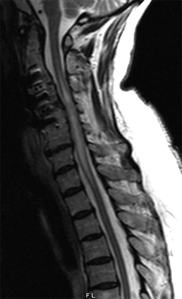
Q7: A 75-year-old woman with history of prior cervical spine decompression presented with progressive neck pain, asymmetric hand tingling, and neuropathic pain, followed by urinary incontinence and gait difficulty. Sagittal T2 weighted imaging likely shows?.
    a: NMO.
    b: Neurosarcoidosis.
    c: Paraneoplastic myelopathy.
    d: Cord ischaemia and oedema.
Q8: Post gadolinium imaging shows?.
    a: Cord infarction.
    b: Transverse myelitis.
    c: Fibrocartilaginous embolism.
    d: Pancake sign.
Q9: The following four questions all relate to patients with acute myelopathy. Axial cross sections of the cord shown. Patient A is likely suffering from ?.
    a: NMO.
    b: MOG antibody disease.
    c: Cord Infarction.
    d: Transverse myelitis.
Q10: Patient with acute myelopathy. Patient B is likely suffering from ?.
    a: NMO.
    b: MOG antibody disease.
    c: Cord infarction.
    d: Transverse myelitis.
Q11: Patient with acute myelopathy. Patient C is likely suffering from ?.
    a: NMO.
    b: MOG antibody disease.
    c: Cord infarction.
    d: Transverse myelitis.
Q12: Patient with acute myelopathy. Patient D is likely suffering from ?.
    a: NMO.
    b: MOG antibody syndrome.
    c: Cord infarction.
    d: Transverse myelitis.
Q13: 45 year old woman with gradually progressive lower extremity numbness, imbalance, and stiffness over 5 months. What is the notable feature on spinal cord imaging?.
    a: Longitudinal extensive T2 hyperintense lesion on sagittal images.
    b: Diffuse hyperintensity on axial T2 weighted images.
    c: Symmetrical enhancement of both lateral columns.
    d: Snake eyes sign.
Q14: What is the likely aetiology of this presentation?.
    a: Vitamin B12 deficiency.
    b: ALS.
    c: Copper deficiency.
    d: Paraneoplastic myelitis .
Q15: Which of the following CSF profiles is most compatible with the patient described in the two previous questions?.
    a: Protein 2.2 g / Lymphocytes 800 / PML 550 .
    b: Protein 1.72 g / Lymphocytes 150 / PML 5 .
    c: Protein 0.5 g / Lymphocytes 40 / PML 2 .
    d: Protein 6 g / Lymphocytes 70 / PML 2 .
Q16: 30 yo female known with HIV, with CD4 count of 35 and progressive myelopathy. CSF showed protein of 2.0 g/l; 82 lymphocytes, 2 PML, glucose 2.9; Gne expert negative; FTA positive (CSF). Chest X Ray suggestive of pulmonary TB.What is the likely cause?.
    a: NMO.
    b: TB myelitis.
    c: MOG antibody disease.
    d: Neurosyphilis.