
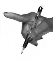
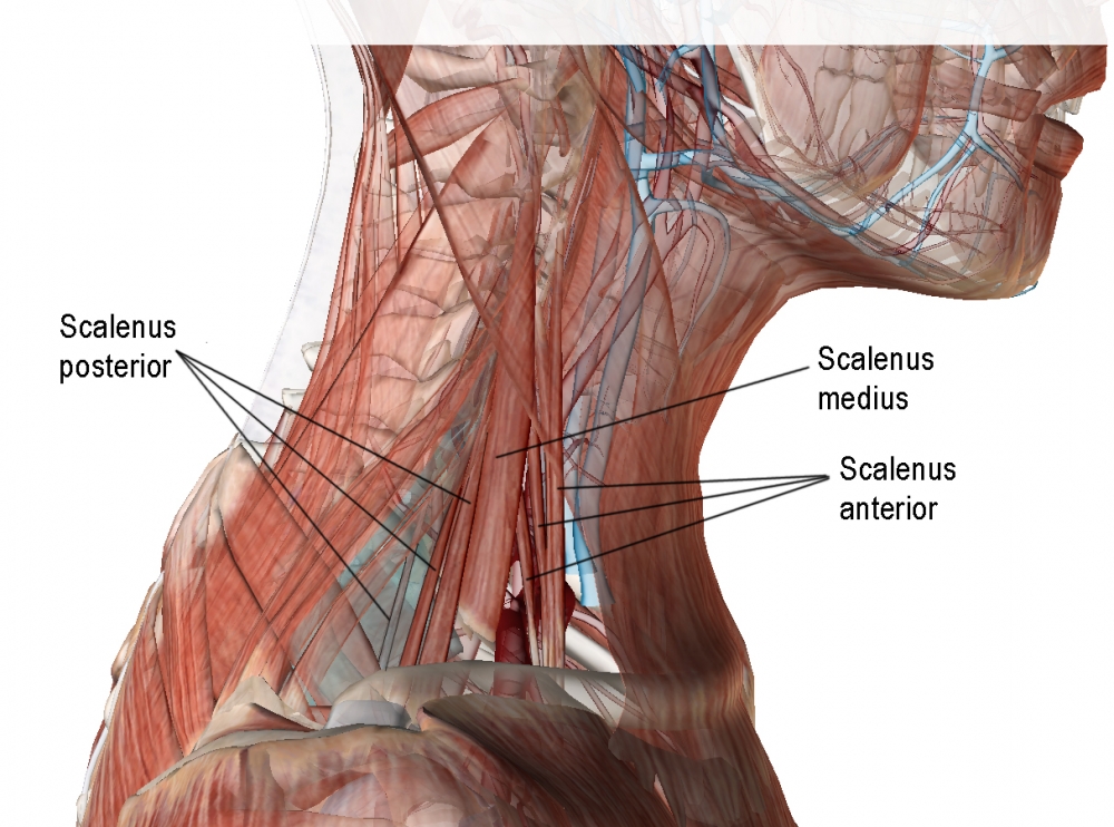
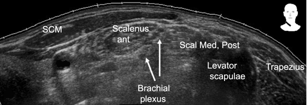
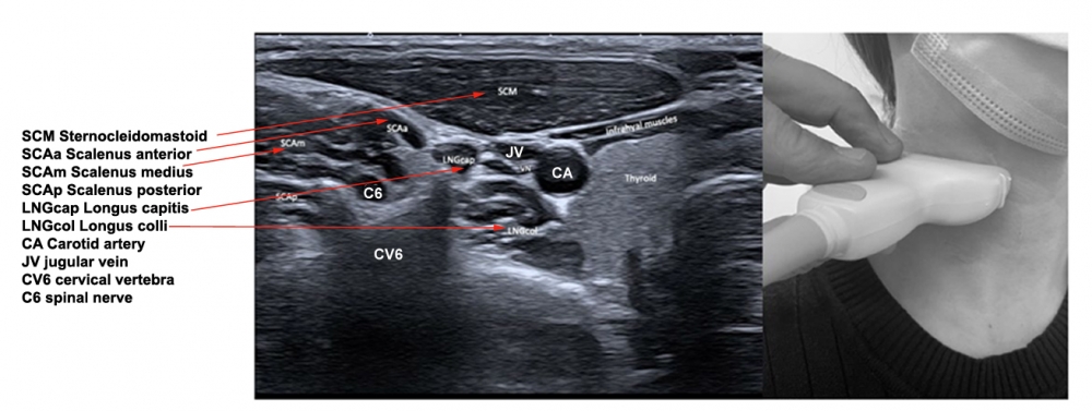
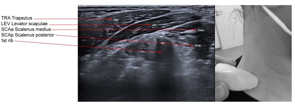
The scalene muscles are identified between the sternocleidomastoid and the levator scapulae. With insertion of the EMG needle into the scalenus muscle, the patient is asked to breathe in as deeply as possible. Since the scalenus is an auxiliary respiratory muscle, EMG activity should be clearly heard on the EMG. Generally either the scalenus posterior or the scalenus medius are treated. Both muscles have the same function: to elevate the rib to which they attach, and to bend and rotate the neck.
Scalenus anterior lies behind the sternocleidomastoid, over the lateral aspect of the neck.
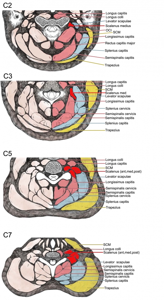
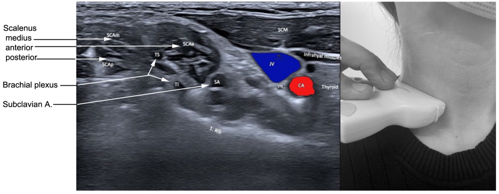
The muscle arises from the anterior tubercles of the transverse processes of C3-C6, and attaches onto the scalene tubercle, on the inner border of the first rib. It is located between the subclavian vein and the subclavian artery. The roots of the brachial plexus pass posterior to it. The phrenic nerve crosses its anterior surface.
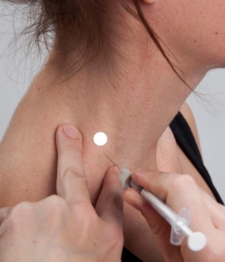
Scalenus medius is the largest muscle of the scalenus group, and arises from the transverse processes of the cervical vertebrae and inserts into the superior portion of the first rib. This muscle can elevate the first rib or bend and rotate the neck. A needle can be placed into the scalenus medius muscle by palpating the belly of the muscle, in the floor of the posterior triangle, two finger breadths anterior to the anterior border of the trapezius muscle. At this point the muscle is just beneath the skin. The upper portion can be inserted by placing the needle just anterior to the lateral edge of the splenius capitis muscle to a depth of 1.5 to 3.0 cm.
Scalenus posterior arises from the posterior portion of the transverse processes of C5–6, passing medially and posteriorly to the scalenus medius, and inserts into the outer surface of the second rib, deep to the attachment of the serratus anterior. The scalenus posterior elevates the second rib or bends and slightly rotates the neck.
| WARNING: neurovascular structures |
-
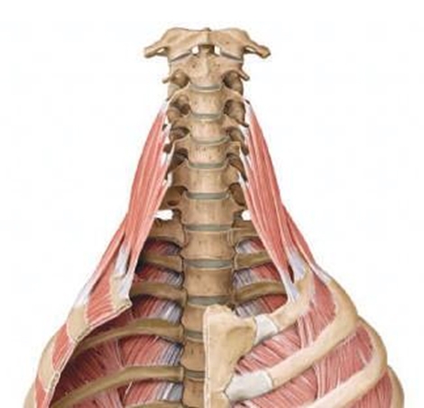
Origin:
Anterior: anterior tubercles of transverse processes of third to sixth cervical vertebrae (C3–6).
Middle: transverse processes of second to seventh cervical vertebrae (C2–7).
Posterior: posterior tubercles of transverse processes of fourth to sixth cervical vertebrae (C4–6). -
Insertion:
Anterior: scalene tubercle and upper surface of first rib.
Middle: upper surface of first rib, behind groove for subclavian artery.
Posterior: upper surface of second rib. -
Action:
Acting on both sides: flex neck; raises first or second rib during active respiratory inhalation.
Acting on one side: tilts neck to the same side, and rotates head.
 |
 |
 |
 |
 |
 |
|
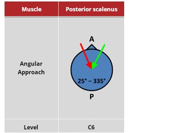
Figure 7. Trajectory of approach and relationships of the surrounding muscles to scalenus
|
|
 |
 |
(vv)Post Scalenus.mp4(tt)
From: Fietzek UM, Nene D, Schramm A, Appel-Cresswell S, Košutzká Z, Walter U, Wissel J, Berweck S, Chouinard S, Bäumer T. The Role of Ultrasound for the Personalized Botulinum Toxin Treatment of Cervical Dystonia. Toxins (Basel). 2021 May 20;13(5):365.
From: Fietzek UM, Nene D, Schramm A, Appel-Cresswell S, Košutzká Z, Walter U, Wissel J, Berweck S, Chouinard S, Bäumer T. The Role of Ultrasound for the Personalized Botulinum Toxin Treatment of Cervical Dystonia. Toxins (Basel). 2021 May 20;13(5):365.
From: Fietzek UM, Nene D, Schramm A, Appel-Cresswell S, Košutzká Z, Walter U, Wissel J, Berweck S, Chouinard S, Bäumer T. The Role of Ultrasound for the Personalized Botulinum Toxin Treatment of Cervical Dystonia. Toxins (Basel). 2021 May 20;13(5):365.