
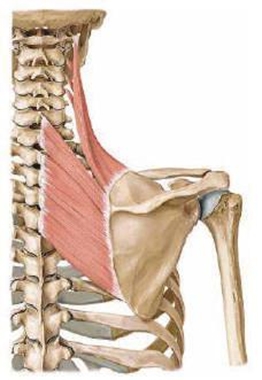
- Origin: posterior tubercles of transverse processes of first,second, third, and fourth cervical vertebrae (C1, 2, 3, 4).
- The transverse process attachment of the levator scapulae is anterior to the transverse process attachment of the splenius cervicis and directly superficial (posterior) to the transverse process attachment of the scalenes.
- Insertion: superior part of medial border of the scapula
- Action: acts mainly on the neck inducing a homolateral rotation and lateroflexion1
The levator scapulae lies directly ventral to the edge of the trapezius. In thin patients, the levator is clearly visible if the treated shoulder becomes lifted.
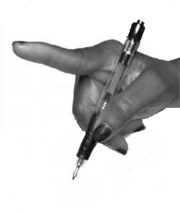
Activation: the patient can be asked to lift the shoulder.
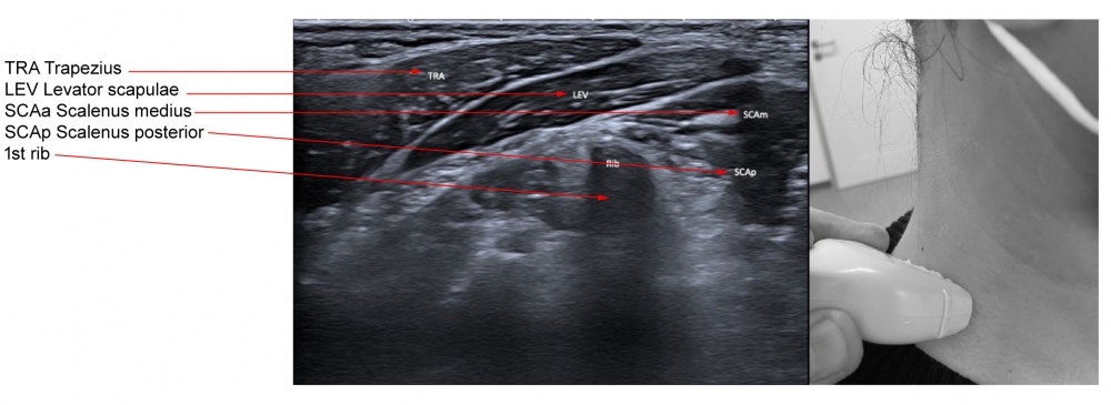
Inferior portion of levator scapulae is covered by trapezius.

Muscle is shown lying deep to trapezius, and immediately ventral to splenius cervicis and splenius capitis.

 |
 |
 |
 |
|
Figure 3. Trajectory of approach and relationships of the surrounding muscles to Levator Scapulae
|
|
 |
 |
(vv)levator scapulae.mp4(tt)
The muscle is typically injected just ventral to the border of trapezius; if there insufficient activation, ask the patient to elevate their shoulder.
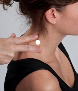
This muscle is organized in four bundles inserted onto the transverse processes of C1, C2, C3 and C4. These bundles are visible in ultrasonographic images and can be injected selectively with botulinum toxin to act at a given vertebral level.
(Note that the transverse process of the atlas (C2) is easily palpable just posterior to the posterior border of the ramus of the mandible, directly inferior to the ear, and directly anterior to the mastoid process of the temporal bone.)
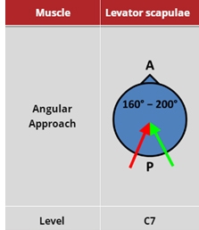
The largest part of the levator scapulae muscle, where the four bundles meet, is located at the muscle's insertion into the medial border of the scapula. Injecting at this site affects the muscle globally and needs a careful approach because of the risk of hitting the pulmonary apex and causing a pneumothorax.
From: Fietzek UM, Nene D, Schramm A, Appel-Cresswell S, Košutzká Z, Walter U, Wissel J, Berweck S, Chouinard S, Bäumer T. The Role of Ultrasound for the Personalized Botulinum Toxin Treatment of Cervical Dystonia. Toxins (Basel). 2021 May 20;13(5):365.

From: Fietzek UM, Nene D, Schramm A, Appel-Cresswell S, Košutzká Z, Walter U, Wissel J, Berweck S, Chouinard S, Bäumer T. The Role of Ultrasound for the Personalized Botulinum Toxin Treatment of Cervical Dystonia. Toxins (Basel). 2021 May 20;13(5):365.