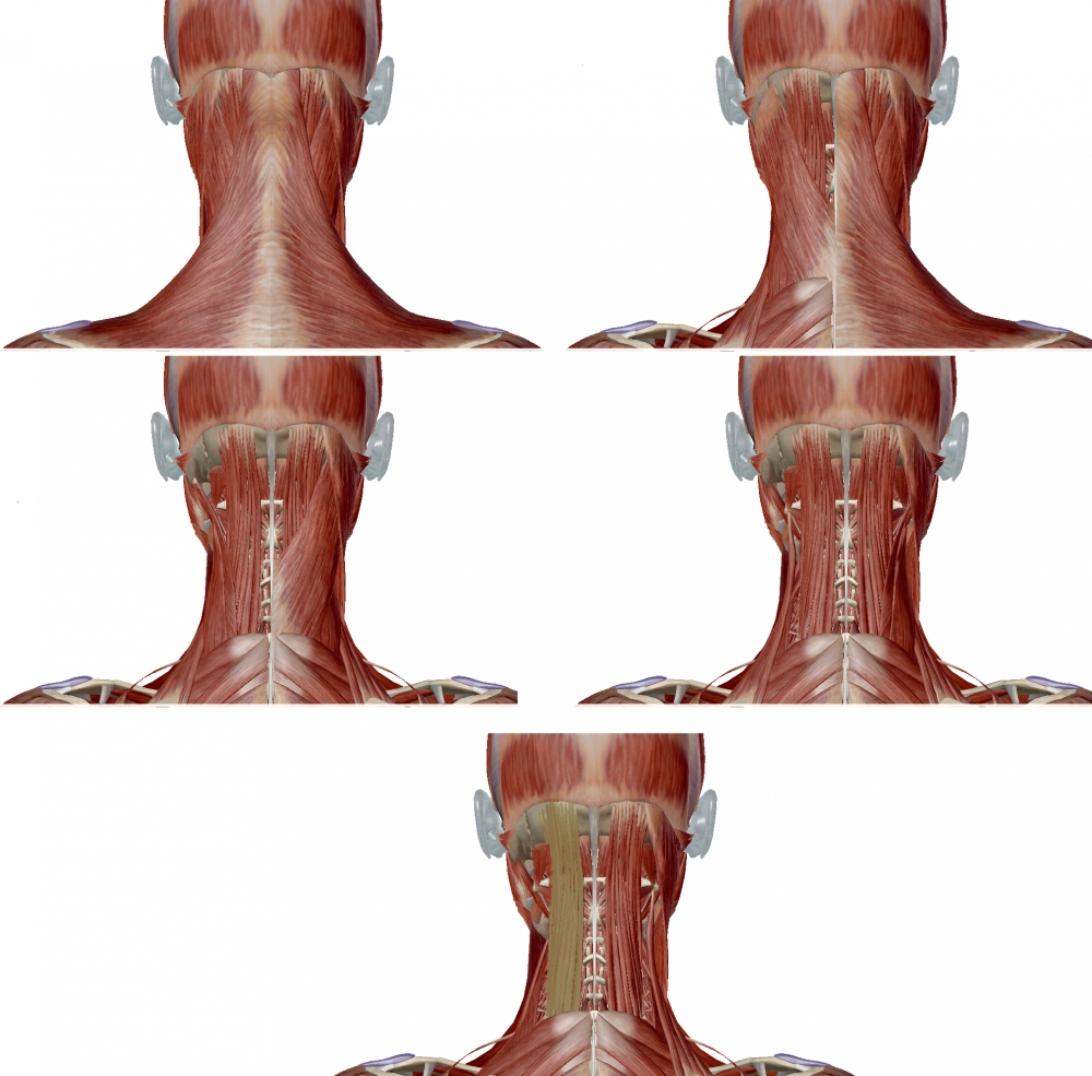
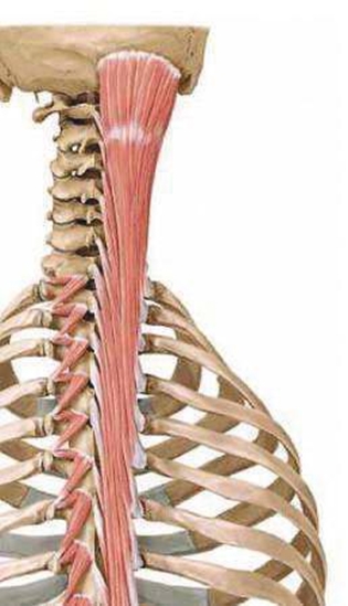
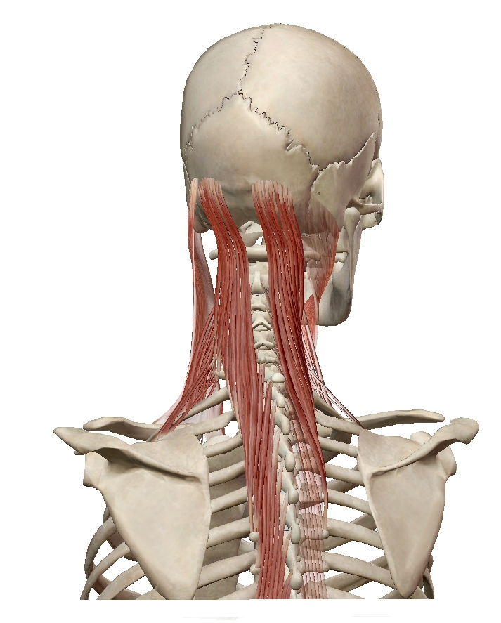
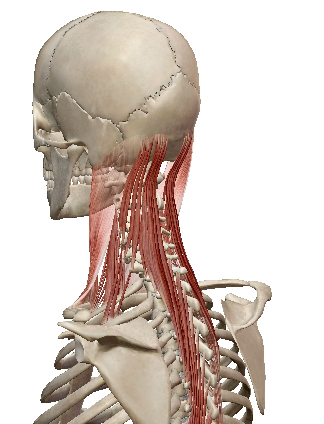
Semispinalis capitis is a long paired muscle that belongs to the deep layer of muscles of the back. It forms the superior, and largest, component of the three-part semispinalis muscle. With semispinalis cervicis and semispinalis thoracis forming the middle and inferior divisions of the muscle, respectively.
The muscle arises from transverse processes of C3-T6, and posterior spinous processes of C3-T1. The muscles inserts between the superior and inferior nuchal lines of the occipital bone.
The semispinalis is covered by the trapezius, below the semispinalis are the paraspinal muscles.
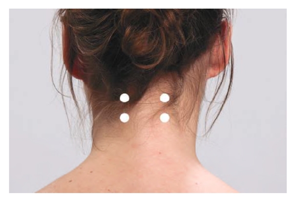
One injection is given one centimeter below the hairline, and the 2nd about 2.5 cm below that, as shown in the figure (Surface Anatomy: Injection Sites). If the neck is flexed (chin towards the chest) the muscle is relaxed and injections less painful; activation of the muscle is achieved by moving the head backwards against the examiner’s hand.
-
Origin: transverse processes of the lower four cervical and upper six or seven thoracic vertebrae (C4–T7).
-
Insertion: between the superior and inferior nuchal lines of the occipital bone.
-
Action: Bilateral contraction leads to extension of the neck.
Unilateral contraction causes contralateral rotation
|
|
 |
 |
 |
 |

From: Fietzek UM, Nene D, Schramm A, Appel-Cresswell S, Košutzká Z, Walter U, Wissel J, Berweck S, Chouinard S, Bäumer T. The Role of Ultrasound for the Personalized Botulinum Toxin Treatment of Cervical Dystonia. Toxins (Basel). 2021 May 20;13(5):365.
(vv)semispinalis capitis.mp4(tt)
1. Trapezius and sternocleidomastoid muscles removed
2. Splenius removed.
3. Longissimus capitis and cervicis removed.
4. Levator scapulae removed showing semispinalis capitis.