-
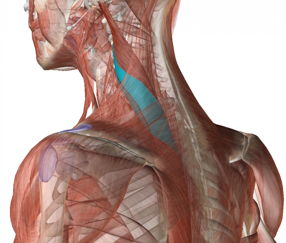
Origin: Transverse processes of the first 5 thoracic vertabrae
-
The transverse process attachment of the levator scapulae is anterior to the transverse process attachment of the splenius cervicis and directly superficial (posterior) to the transverse process attachment of the scalenes.
-
Insertion: Posterior tubercles of the transverse processes of C2-C5
-
Action: Bilateral contraction leads to extension of the neck.
Unilateral contraction causes lateral flexion and rotation of the head ipsilaterally
This muscle is deep to the levator scapulae muscle's attachment to the transverse processes, and its depth greatly varies from patient to patient, being often unexpectedly superficia in some adults.
Ultrasound Technique
1. Palpate the spinous process of the axis and move the operator's finger to the third cervical (C3) spinous process.
2. Place the linear transducer horizontally at this level over the triangle formed posteriorly by the trapezius, anteriorly by the sternocleidomastoid, and inferiorly by the middle third of the clavicle.
3. Move the transducer a little dorsally and localize the transverse process of C3 on the right lower part of the screen. The longissimus capitis muscle can be seen in the direction of 10â€11 o'clock from the transverse process with a deformed and widened letter Dâ€shape (the vertical line of the letter D represents the fascia, discriminating it from the semispinalis capitis
muscle)
4. After the injection, repeat the procedure at the C5 level. Color Doppler imaging at this level occasionally detects an artery, which is the superficial branch of the transverse cervical artery. .

From: Fietzek UM, Nene D, Schramm A, Appel-Cresswell S, Košutzká Z, Walter U, Wissel J, Berweck S, Chouinard S, Bäumer T. The Role of Ultrasound for the Personalized Botulinum Toxin Treatment of Cervical Dystonia. Toxins (Basel). 2021 May 20;13(5):365.