
See also: Long Segment Myelopathy
LOCATION |
SAGITTAL T2 |
POST CONTRAST SAGITTAL |
OTHER FEATURES |
|---|---|---|---|
|
Commonly, Cervical1> Thoracic |
Spinal cord oedema is typically seen in the acute phase of MS myelitis, |
Enhancement is present in most acute lesions; |
Spinal Cord atrophy correlates with |
|
|
Image from Ref 1
Image from Ref 3 |
Image from Ref 3 |
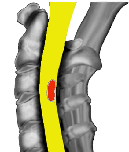
Typical apple core lesion of chronic MS1.
|


|
COMPARISON OF MOG, NMO AND MS |
AXIAL T2 (NON CONTRAST) |
|---|---|
|
MOG: Sagittal T2-hyperintense line (white arrowhead) surrounded by hazier T2 hyperintensity (yellow arrowheads). |
Lesions are typically peripheral, and wedge-shaped or |
 |
|


